Cervical Acceleration-Deceleration Injury

Whiplash & Spinal Trauma

Whiplash is among the most misunderstood and underdiagnosed injuries in medicine. Symptoms are frequently delayed, standard imaging routinely misses the underlying ligament damage, and untreated injuries can progress to permanent spinal instability. Dr. Moore specializes in the objective evaluation and treatment of these injuries.

72h
Typical symptom onset delay
0
Ligament signal on standard X-ray
1 of 1
Digital Motion X-Ray in Mobile, AL

Understanding the Injury

What Actually Happens During a Whiplash Event

Whiplash — formally termed Cervical Acceleration-Deceleration (CAD) injury — describes the rapid, forced flexion-extension of the cervical spine that occurs during a sudden impact. The mechanics unfold in milliseconds, well beyond the body's reflexive muscular protection response.

During a rear-impact collision, the torso is accelerated forward by the car seat while the head, unsupported, initially remains stationary. This differential creates a shearing force across the cervical spine. The lower cervical segments hyperextend first, while the upper segments simultaneously flex — creating an S-shaped curvature that places the spinal discs, facet joints, and ligaments under extreme tensile and compressive load.

The entire biomechanical event typically takes 150–300 milliseconds. Human reaction time is approximately 200 milliseconds. This means the injury sequence is complete before the body's muscles can contract to stabilize the neck. The passenger's head-restraint position, seat stiffness, awareness of the impending impact, and the direction of force all influence the degree of injury.

Key fact: Research has documented significant cervical injury at impact speeds as low as 5–8 mph. Occupants of vehicles with little visible damage may sustain substantial soft-tissue injury because energy-absorbing bumpers reduce structural damage to the vehicle while transferring force directly to the occupants.

The joints, discs, ligaments, and musculature of the neck all absorb this energy in varying proportions depending on individual anatomy, the direction of force, and whether the occupant was braced at the moment of impact.

Why Symptoms Are Delayed

Feeling Fine After an Accident Does Not Mean You Are Fine

It is common — and clinically significant — for whiplash symptoms to be absent or minimal immediately after a collision. This delay leads many patients to forgo evaluation, a decision that can have long-term consequences.

The Neurochemical Response

At the moment of impact, the body releases a cascade of stress hormones — primarily epinephrine (adrenaline) and cortisol. These hormones suppress pain perception as part of the fight-or-flight response, allowing an injured person to function in the immediate aftermath of trauma. This neurochemical masking typically persists for hours to days.

The Inflammatory Cascade

Soft tissue injury triggers an inflammatory response that builds over 24–72 hours following trauma. Pro-inflammatory cytokines accumulate in damaged tissue, nerve endings become sensitized, and edema develops around injured structures. Most patients report peak pain and stiffness 2–3 days post-accident — precisely when inflammation is at its height.

Clinical implication: Waiting until symptoms are severe to seek evaluation can complicate causation documentation and allow injury patterns to become more difficult to treat. Early evaluation — even within the first 24 hours when symptoms are minimal — establishes a clear clinical record of findings that correlates with the accident event.

The Protective Muscle Spasm Response

Injured cervical structures trigger involuntary muscle guarding — a protective splinting response that maintains spinal position and reduces the immediate pain signal. As days pass and the nervous system recalibrates, spasm patterns shift, and pain referral patterns emerge to the shoulders, arms, and head. What begins as neck stiffness may evolve into headaches, radiating arm pain, cognitive changes, and sleep disturbance.

Neurological Adaptation Over Time

Persistent injury to joint mechanoreceptors — the sensory structures within spinal ligaments that inform the brain about head position and movement — can lead to proprioceptive deficits, dizziness, visual disturbances, and difficulty concentrating. These findings, sometimes dismissed as psychological, have well-documented neuroanatomical explanations in the whiplash literature.

Spinal Anatomy in Motion

The Stages of Whiplash Injury

Understanding what happens to the cervical spine during a whiplash event — and what the long-term consequences of untreated injury look like — helps clarify why objective evaluation is essential.
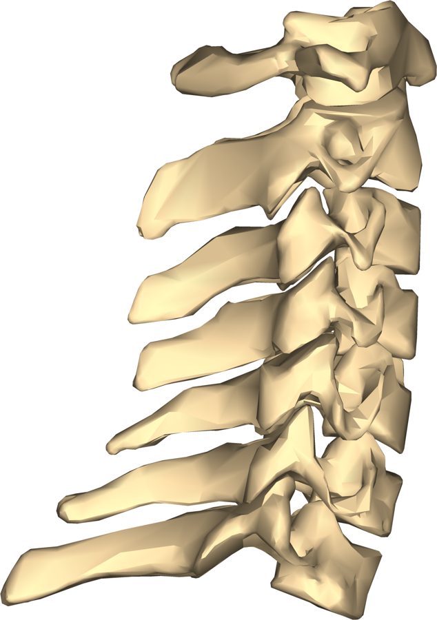
Lateral view of the cervical vertebrae

01 — Normal Anatomy

Healthy Cervical Spine

The normal cervical spine maintains a gentle lordotic (forward) curve of approximately 35–45 degrees. Seven vertebrae are separated by hydrated discs acting as shock absorbers. Paired facet joints, interspinous ligaments, and the anterior/posterior longitudinal ligaments provide stability while allowing full range of motion.

Illustration: Anatomography, CC BY-SA 2.1 JP
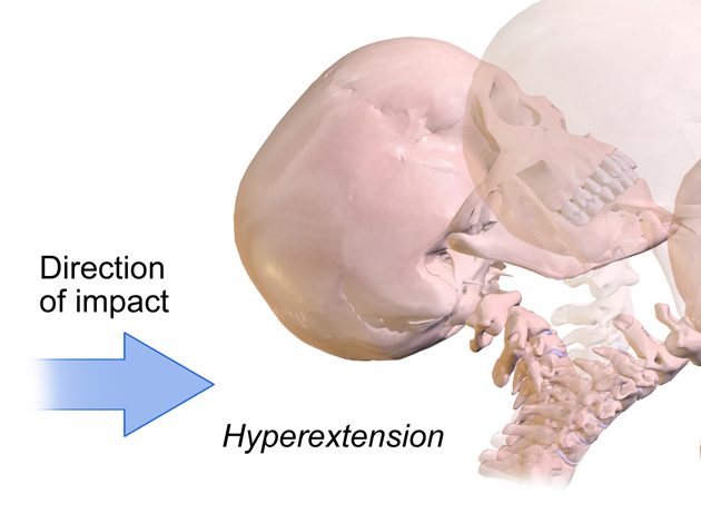
Illustration of hyperextension of the neck during a rear-impact collision

02 — Phase One

Hyperextension Phase

As the torso is thrust forward, the lower cervical spine is forced into extension while the upper segments flex — producing an abnormal S-curve. The posterior elements (facet joint capsules, posterior ligaments, joint capsules) are placed under rapid tensile load, often exceeding the elastic limits of the ligament fibers. This phase occurs in approximately 50–75 milliseconds.

Illustration: BruceBlaus, CC BY-SA 4.0, cropped and adapted
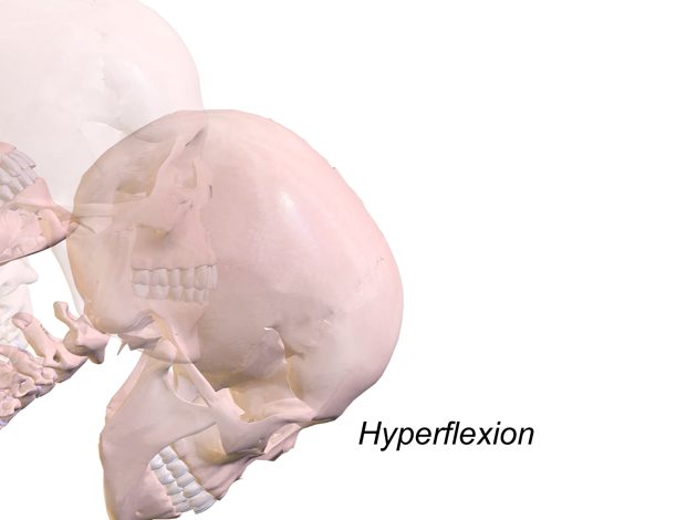
Illustration of the hyperflexion rebound phase of whiplash

03 — Phase Two

Hyperflexion Rebound

Following the initial extension, the head rebounds forward into hyperflexion, loading the anterior structures — the anterior longitudinal ligament, disc annulus, and anterior disc space. This rapid reversal can produce disc tears and anterior ligament damage while re-straining the posterior structures already injured during phase one. The total event lasts 150–300 milliseconds.

Illustration: BruceBlaus, CC BY-SA 4.0, cropped and adapted
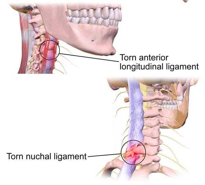
Illustration of a torn anterior longitudinal ligament and torn nuchal ligament in whiplash

04 — Ligament Injury

Ligament Laxity & Instability

Spinal ligaments — the anterior and posterior longitudinal ligaments, interspinous ligaments, and joint capsule ligaments — have limited blood supply and do not heal with the same tensile strength as muscle. Partial tears can heal with residual laxity — a reduced ability to resist normal vertebral motion. This instability may not be visible on static X-ray and is better assessed with motion imaging such as flexion-extension radiographs or Digital Motion X-Ray.

Illustration: BruceBlaus, CC BY-SA 4.0, cropped and adapted
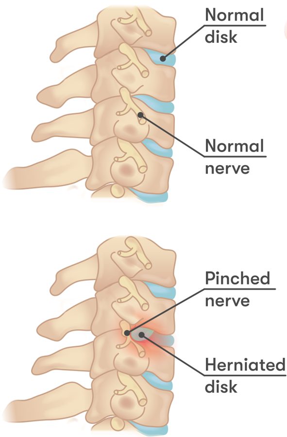
Comparison of a normal cervical disc and a herniated disc pinching a nerve

05 — Disc Pathology

Intervertebral Disc Involvement

The intervertebral disc consists of a fibrous outer ring (annulus fibrosus) surrounding a gel-like center (nucleus pulposus). Rapid compressive and tensile forces during a whiplash event can create annular tears, allowing nucleus material to migrate and press against the exiting nerve root — producing radiating pain, numbness, or weakness in the arm or hand corresponding to the affected cervical level.

Illustration: InjuryMap, CC BY-SA 4.0, cropped and adapted
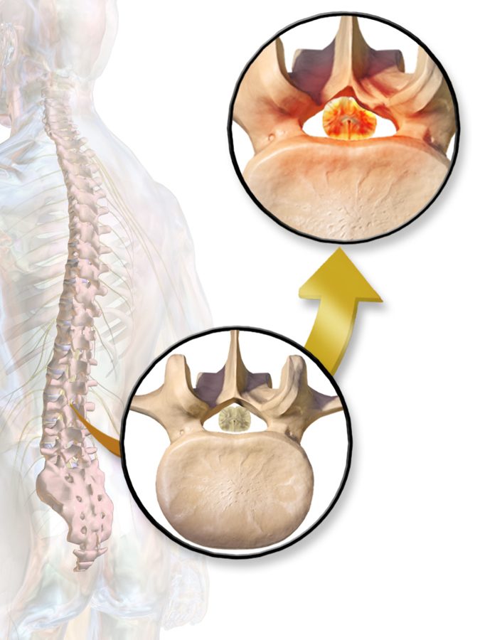
Illustration of spinal canal narrowing in spinal stenosis

06 — Long-Term Consequences

Chronic Degeneration If Untreated

Spinal instability is associated in the literature with a cascade of degenerative change. Abnormal segmental motion produces chronic inflammation in the facet joints, accelerates disc desiccation and collapse, and stimulates bone spur formation. Over years, this process can contribute to narrowing of the spinal canal (stenosis) and foraminal narrowing that compromises nerve root space — which is why early, appropriate evaluation matters.

Illustration: Blausen Medical, CC BY 3.0 (general illustration; lumbar spine shown)

Clinical Definitions

The Difference Between Sprains and Strains

These terms are frequently used interchangeably, but they describe injuries to fundamentally different tissues with distinct prognoses and implications for long-term stability.

Ligament Injury

Sprain

  • Injury to ligaments — the fibrous bands connecting bone to bone
  • Ligaments stabilize joints and resist excessive motion
  • Limited vascularity means slow and often incomplete healing
  • Partial tears may heal with residual laxity (stretch injury)
  • Instability may not be symptomatic until progressive degeneration occurs
  • Graded I (stretch), II (partial tear), III (complete rupture)
  • Best assessed with Digital Motion X-Ray or MRI

Muscle / Tendon Injury

Strain

  • Injury to muscles or their tendinous attachments (muscle-to-bone)
  • Muscles generate movement and provide dynamic stability
  • Well-vascularized tissue; generally heals within 6–12 weeks
  • Results in pain, spasm, and temporarily reduced strength
  • Healed muscle returns to near-normal tensile strength with rehabilitation
  • Graded I (microtear), II (partial tear), III (complete rupture)
  • Assessed with palpation, MRI, and functional testing

Clinical significance: A patient who sustains a Grade II cervical ligament sprain (partial tear) may recover from the overlying muscle strain within a few weeks and feel substantially better — while the underlying ligament laxity creates silent instability that drives long-term degenerative change. This is why objective imaging of ligament integrity, not just symptom resolution, is the appropriate endpoint for whiplash evaluation.

Motion Imaging for Ligament Evaluation

Why Standard X-Ray Misses
the Most Important Findings

Ligaments are soft tissue structures — they contain no calcium and produce no signal on standard radiography. A standard cervical X-ray in a whiplash patient may appear completely normal while the stabilizing ligament system is severely compromised.

STATIC X-RAY Ligaments: not visible DIGITAL MOTION X-RAY Laxity Instability quantified

Real-Time Fluoroscopic Video

DMX captures continuous X-ray imaging at 30 frames per second while the patient performs guided cervical movements. The result is a video of the spine in motion, revealing dynamic instability that no static image can show.

Quantified Instability

Abnormal vertebral translation — the hallmark of ligament laxity — is measured in millimeters. Published radiological standards and the AMA Guides define thresholds for abnormal motion, providing objective, measurable data.

Only Available DMX in Mobile, AL

Optimum Chiropractic Care operates the only Digital Motion X-Ray unit in the Mobile, Alabama region. Studies are performed on-site.

Medico-Legal Documentation

DMX findings are recorded as video with measured values, so they can be reviewed by other clinicians. Dr. Moore provides structured reports of the findings when documentation is needed.

Have You Been in an Accident?

Do not wait for symptoms to worsen. Early evaluation provides the clearest clinical picture and an accurate record of your findings close to the time of injury. Dr. Moore accepts new patients throughout the Mobile Bay area.

Request an Evaluation